.png)
.png)


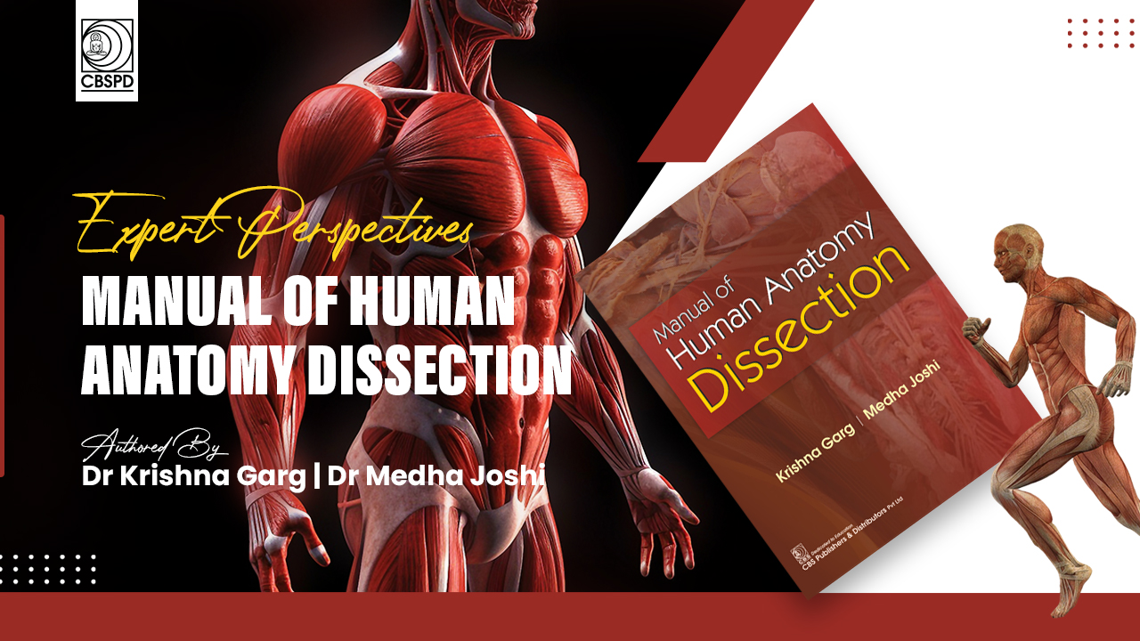
Expert Perspectives: Manual of Human Anatomy Dissection
.jpg)
Dr Karishna Garg| Dr Medha Joshi
“I learn anatomy not from books but from dissections, not from the tenets of philosophers but from the fabric of nature”.
-William Harvey
Dissection is but one of the means of learning anatomy. Dissection of an animal is the learning of zoology, while dissection of a human cadaver is the learning of anatomy.
Dissection is a time-tested means of learning anatomy for centuries despite the technological advances. Dissection is a very specialised scientific procedure and is different from an "autopsy" or "postmortem".
During the anatomical dissection, the medical student, under the instructions of an experienced teacher, works slowly and steadily, identifying various structures. As of now, it takes almost one year during the preclinical course to dissect the whole body and there is a definite procedure to perform the dissection of any region. For this, we have some Indian books which give steps of dissection with the hand-drawn/computer-drawn diagrams taken from textbooks of anatomy and are not the actual dissection photographs/ diagrams; defeating the whole exercise of dissection. A few foreign atlases are available with beautiful dissection photographs but these look "too good to be true" for our milieu.
Mr SK Jain, Chairman and Managing Director, CBS Publishers & Distributors, proposed a "Dissector of anatomy". After many deliberations, considering the real requirements of MBBS students, we decided to give the entire dissection in one volume only. The highlight of the volume would be to provide "actual ordinary dissection photographs" as seen in the dissection halls of a medical college.
There have been a multitude of hurdles in obtaining the "actual dissected specimens" from various sources. We could have taken specimens from any good museum, but these would be "too real for actual ordinary dissection specimen" as museum specimens are the artwork of senior teachers and technicians.
The first group of teachers to help us in this difficult and tough endeavour was the Principal, Rama Medical College, Kanpur. Later, assistance came from Bakson Homeopathic Medical College and Hospital, Greater Noida, UP. We have emphasised on the dissection photographs, not on illustrations giving only theoretical knowledge.
Actual dissection specimens were also available from DDU National Institute for Persons with Physical Disabilities, New Delhi, where we performed the dissection ourselves and took requisite photographs. Some photographs were also taken from Subharti Medical College, Meerut.
Towards the end of three years' ongoing struggle, AllMS-Raipur pitched in, giving us the remaining photographs as the work on the Manual got completed. Despite all these efforts, a few diagrams are still hand- or computer-drawn, which we will like to replace in the future editions.
The aim of this dissector is to reach, locate and identify the structures in a particular region. Their description is beyond the scope of this book and has to be learnt from a standard Textbook of Anatomy, BD Chaurasia's Human Anatomy, 11th edition, Volume 1 to 4. It has been done deliberately to limit the size of the book, avoiding repetition of theoretical text material. Thus, this Manual on dissection is a supplement to the Textbook of Anatomy. It should be able to provide holistic, useful, photographic memory for students’ long, happy clinical years.
Steps of dissection are given in a simplified way with as many "ordinary dissection photographs" as possible to make the whole exercise easy and interesting. It will make many senior students or doctors nostalgic about their days in the dissection hall.
In the majority of medical colleges, there is a shortage of cadavers and scarcity of time. In most of the medical institutions, there are 20-30 students for one region being dissected and the time allocated to the study of anatomy is hardly one year.
In this dissector, the text of dissection is divided into Introduction and six sections
· Thorax
· Lower Limb
· Abdomen and Pelvis
· Head and Neck
· Brain
The chapter on Introduction relates to getting the cadaver, the embalming procedure, maintaining and caring for the cadaver, and finally disposing its parts. It also includes various instruments for dissection.
Every section is divided into various chapters. Each chapter starts with "learning objectives" and "overview ", providing the bird's-eye view of the particular topic. These are followed by "steps of dissection" and actual photographs of the concerned area. Emphasis has been given on the structures visible only.
The chapter ends with a few viva voce examination questions to help in relearning the topic. This procedure is followed in all chapters of a section. At the end of each section, 10-15 spots are given. These are important from the examination point of yiew. This is the simple procedure followed in the present book.
The only authentic way to learn anatomy is to draw, draw and draw coloured diagrams and marking structures on your body.
We are reminded of the quotation:
“If I hear, I forget,
If I see, I remember
and if I do, I understand”.
Wishing you happy understanding and learning of anatomy!
From the Desk of CBS Publishers and Distributors
The following books are now available on our website www.cbspd.com.
1. Manual of Human Anatomy Dissection
2. The All-new and updated edition of BD Chaurasia’s Human Anatomy now comes in 4 volumes, each dedicated to:
·Volume 1: BD Chaurasia’s Human Anatomy: Upper Limb|Thorax
Volume 2: BD Chaurasia’s Human Anatomy: Lower Limb|Abdomen and Pelvis
Volume 3: BD Chaurasia’s Human Anatomy: Head and Neck
Volume 4: BD Chaurasia’s Human Anatomy: Brain|Neuroanatomy
All the volumes are also available nationwide through all CBSPD dealers. For online purchases, the books have been shelved with Amazon. For any further information/queries about the books, we are happy to assist you via call/WhatsApp on 9599779677.